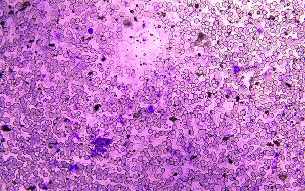
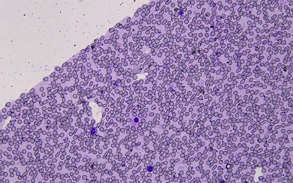
In routine blood examinations, microscopic examination is an examination method that inspectors should master.
In recent years, with the continuous development of science and technology, blood cell analyzers have been widely used in routine blood clinical examinations, but microscopy is still indispensable, otherwise it may add unnecessary trouble to clinical diagnosis, such as misdiagnosis and missed diagnosis.
The following is a blood slice under the MSHOT biological microscope-conventional blood morphology observation (mainly to photograph blood morphology and tissues and observe the count of white blood cells):

The MSHOT biological microscope ML31 is used in the laboratory laboratory of the hospital. This microscope adopts an infinite optical system, which can realize bright field, dark field, and phase contrast multi-function microscopic observation. The focusing system often uses coaxial coarse and fine focusing mechanism, mechanical-style movable stage, stage size 210*140mm, moving range 76*50mm, external 1x digital camera adapter, real-time display of camera field size and eyepiece field of view.
