Mycobacterium tuberculosis is a slender and slightly curved bacillus, sometimes branched. Due to the effects of aging and anti-tuberculosis drugs, it can appear in many forms, such as spherical, beaded or filamentous. Mycobacterium tuberculosis is a typical acid-fast bacillus and is usually difficult to color. It is not easy to be decolorized by hydrochloric acid alcohol after dyeing with aniline dye under antipyretic conditions, which is the acid resistance of bacteria. Stained with the fluorescent dye auramine "O", the bacteria are golden yellow under a fluorescent microscope.
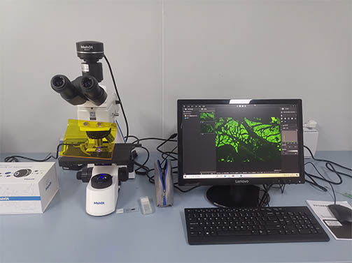
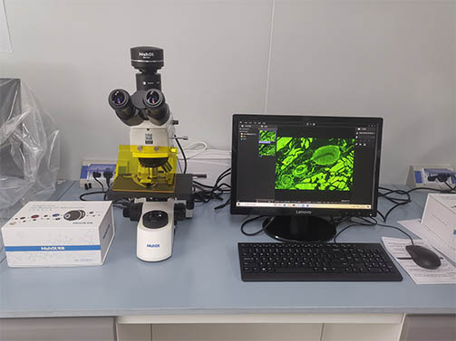
MSHOT fluorescence microscope MF31 was introduced into Inner Mongolia Disease Control for tuberculosis detection. This fluorescence microscope adopts infinity plan achromatic objective and wide-field eyepieces. It can be observed with binocular or trinocular, and is equipped with LED fluorescence excitation device to realize bright field and fluorescence. It is used in clinical laboratory to observe small cells, tissues and other samples.

In order to facilitate image storage and comparative analysis, we have equipped it with a set of 6.3 megapixel resolution microscope cameras. The microscope camera MS60 can better observe and compare the samples, and obtain the same image effect as under the eyepieces, showing true and reliable Renderings. Make the experiment more effective and faster, reduce the time spent on shooting, and greatly improve work efficiency. It can clearly see every detail of the sample. It can see the bright field and fluorescence, so as to achieve better applications.
