

Zebrafish: application of model organism
Zebrafish (Danio rerio) is a common and commonly used model organism. Because zebrafish and human gene have 87% high homology, they are often used to study vertebrate animal development and gene function. Due to the convenience of zebrafish breeding, short breeding cycle, large amount of eggs, in vitro fertilization, in vitro development and transparent embryo body, researchers can observe the fine structure of its tissue and cells, and the overall situation of the sample. It can reveal the molecular mechanism of embryo and organ development, build various human disease and tumor models, establish drug screening and treatment research platform, establish toxicology and aquaculture breeding models, and study and solve major problems in Environmental Science and agricultural science. Progress has been made in the fields of developmental biology, oncology, toxicology, reproductive medicine, teratology, genetics, neuroscience, environmental science, cytology, regenerative medicine and evolutionary theory.
How to see the subtle structural differences in zebrafish? What kind of microscope and microscope camera does it need?

Stereo-fluorescence microscope MZX81 and microscope camera MS23 allow you to clearly observe the structural differences.
Zebrafish development
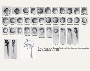
Embryo development is rapid. It takes only 24 hours from the fertilized egg to the complete embryo. 3-5 days after fertilization, every fish can swim and feed freely. The internal organs similar to human body have been built. The body is transparent, zebrafish in the first 7 days of development body transparent, can directly observe the internal organs. Combined with in vivo dye, antibody, nucleic acid probe and other methods, we can observe free or fixed zebrafish in vivo samples. This direct observation has laid a solid foundation for automatic drug screening and drug target organ identification. Among known organisms, fish is a class with acquired immune system.
Cancer research
Zebrafish are used to produce transgenic models for cancer research, including melanoma, leukemia, pancreatic cancer and hepatocellular carcinoma. Zebrafish models expressing mutated BRAF or NRAS oncogenes develop melanoma in the absence of tumor protein (p53). Histologically, these tumors are highly similar to human diseases, are implantable and exhibit a wide range of genomic changes. In another study, the first mock exam was used to study the function of the amplified and overexpressed genes in human melanoma. Setdb1 gene can significantly accelerate tumor formation in zebrafish, which shows its nature as a carcinogen. This is particularly important because setdb1 is known to be involved in epigenetic regulation, and epigenetic regulation is increasingly considered to be the core of tumor cell biology.
Gene expression
Because of its short life cycle and strong controllability, zebrafish is often used as a model animal for genetic research. Knockdown and modified RNA splicing with antisense morpholino are common reverse genetics techniques. Synthetic high molecular morpholine oligonucleotides contain the same nucleosides as DNA and RNA; by combining with complementary sequences, they can reduce the expression of specific genes or hinder the process of other RNA.

Drug research and development
As demonstrated in many ongoing research projects, the zebrafish model enables researchers not only to identify genes that cause human diseases, but also to develop new therapeutic agents in drug development projects. Zebrafish embryo is a rapid, cost-effective and reliable teratogenicity test model. Drug screening with zebrafish can identify new compounds with biological efficacy, or discover new uses of known drugs. For example, a commonly used agent (rosuvastatin) has been found to control the growth of pre adenocarcinoma in zebrafish. At present, a number of small molecule screening have been carried out, some of which have been clinical trials.
Environmental monitoring
Researchers have modified zebrafish's genes to allow it to be used to detect estrogen contamination in water, which is thought to be linked to male infertility. The researchers cloned estrogen sensitive genes and injected them into the fertilized eggs of zebrafish. The resulting transgenic fish turned green when they perceived pollution.
Immune system
In the study of acute inflammation, researchers have established zebrafish models for inflammation research and related processing mechanisms, so that researchers can refine the genetic control mechanism of inflammation, and it is possible to identify potential new drugs. Zebrafish is a widely used model organism in the study of vertebrate innate immunity (innate immunity can phagocytize within 28-30 hours after fertilization, and phagocytosis is an important part of immune response). In contrast, adaptive immunity (also known as specific immunity, acquired immunity and acquired immunity) can reach functional maturity at least four weeks after fertilization.

Regeneration ability
Zebrafish can regenerate the hair cells of fin, skin, heart, lateral line and brain. In 2011, the British Heart Foundation advertised its plan to apply this ability to the human body.
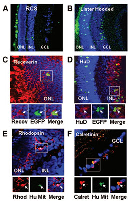
Zebrafish have also been found to regenerate photoreceptors and retinal nerves after trauma. Current studies have shown that this is mediated by the dedifferentiation and proliferation of Muller glia cells. Researchers cut off the caudal fin on the back and abdomen, and analyzed its regeneration to observe its mutation. It has been found that histone demethylase (histone methylation) appears at the amputation site, which makes zebrafish cells reactivate to a renewable state similar to stem cells. In 2012, a study published by Australian scientists showed that zebrafish use a specific protein called fibroblast growth factor to ensure that their spinal cord can heal without glial scar. In addition, hair cells in the lateral line of the posterior body of zebrafish were found to regenerate after trauma or developmental interruption. The study of gene expression during regeneration has enabled the identification of several important signaling pathways, such as Wnt signaling pathway and fibroblast growth factor.
Retinal injury
Another remarkable feature of zebrafish is that it has four kinds of cone cells, in addition to the red, green and blue sensitive cone cells in human body, it also has ultraviolet sensitive cells. Zebrafish can therefore see a very wide spectrum. Therefore, zebrafish is also used to study the development of retina, especially how cone cells form mosaic in retina. Zebrafish and some teleost fishes have attracted much attention because of the mosaic arrangement of cone cells in the retina. Researchers at University College London have developed adult zebrafish stem cells that are found in zebrafish and mammalian eyes and develop into retinal nerves. These cells can be injected into the eye to treat diseases that damage the retinal nerves, covering most eye diseases, including macular degeneration, glaucoma, diabetes related blindness, and so on. The researchers studied m ü ller cells in people's eyes, ranging in age from 18 months to 91 years. In the study, researchers were able to grow these cells into all kinds of retinal nerve cells. The team can easily grow these cells in the laboratory, and transplant the stem cells into the retina of rats to observe the peripheral nerves. The researchers say the stem cells are trying to develop in the same way as they do in humans.

Experiment completed: high quality performance images
Functional imaging is one of the most demanding tasks in the laboratory. MSHOT stereo-fluorescent solutions is favorable.
Completely based on the microscope digital imaging software operation and control ability.
Real time fluorescent multichannel synthesis.
Image measurement.
Camera control.
Image acquisition.
The blackboard on the base of the stereo fluorescence microscope is used to eliminate the scattered light and realize the functional imaging of the sample.
