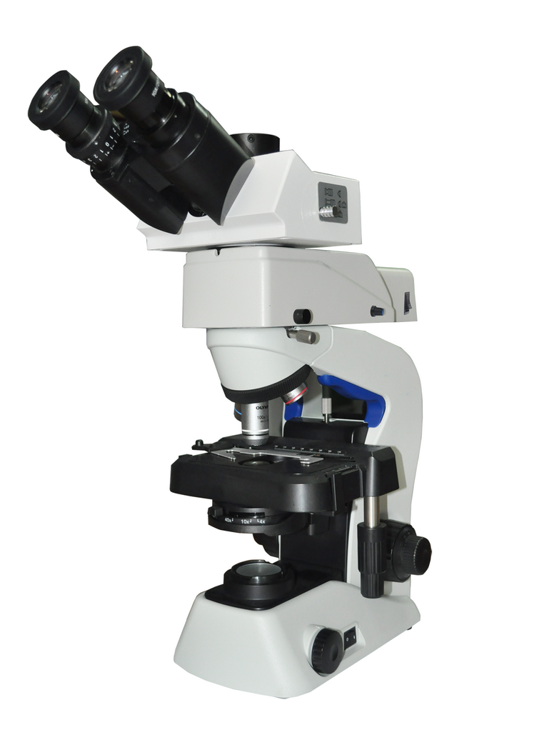
Fungus diagnose is one of the ideal application from LED fluorescence microscope, which takes use of UV excitation group. Hypha is a tubular single filamentous structure of fungus, also a structure of unrelated actinomycetes. Although it is the structural unit of most fungi, most of the mycelium of edible fungus is no poison. With the emergence of the mycelium in the field of medicine, the new market of fungi is gradually formed.
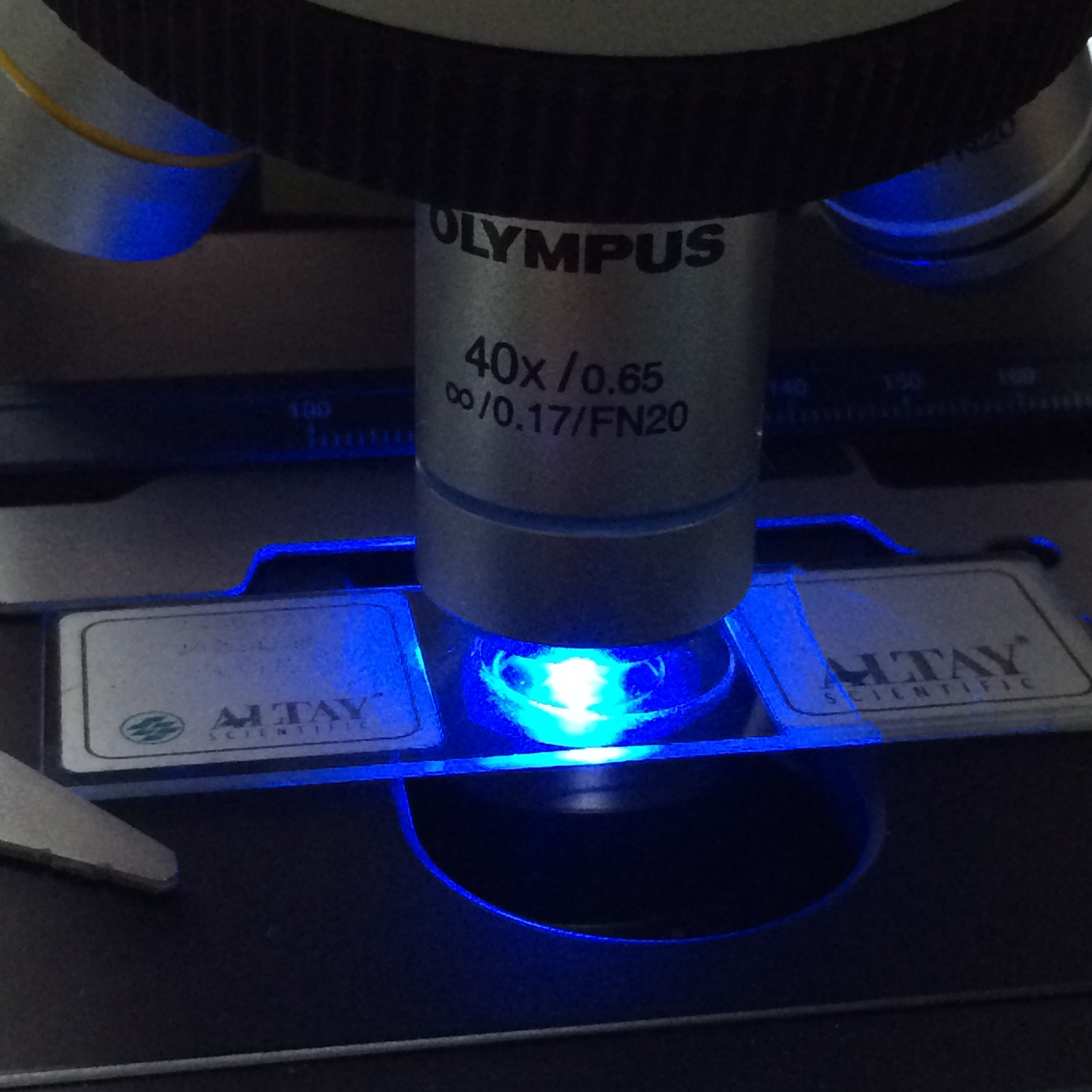
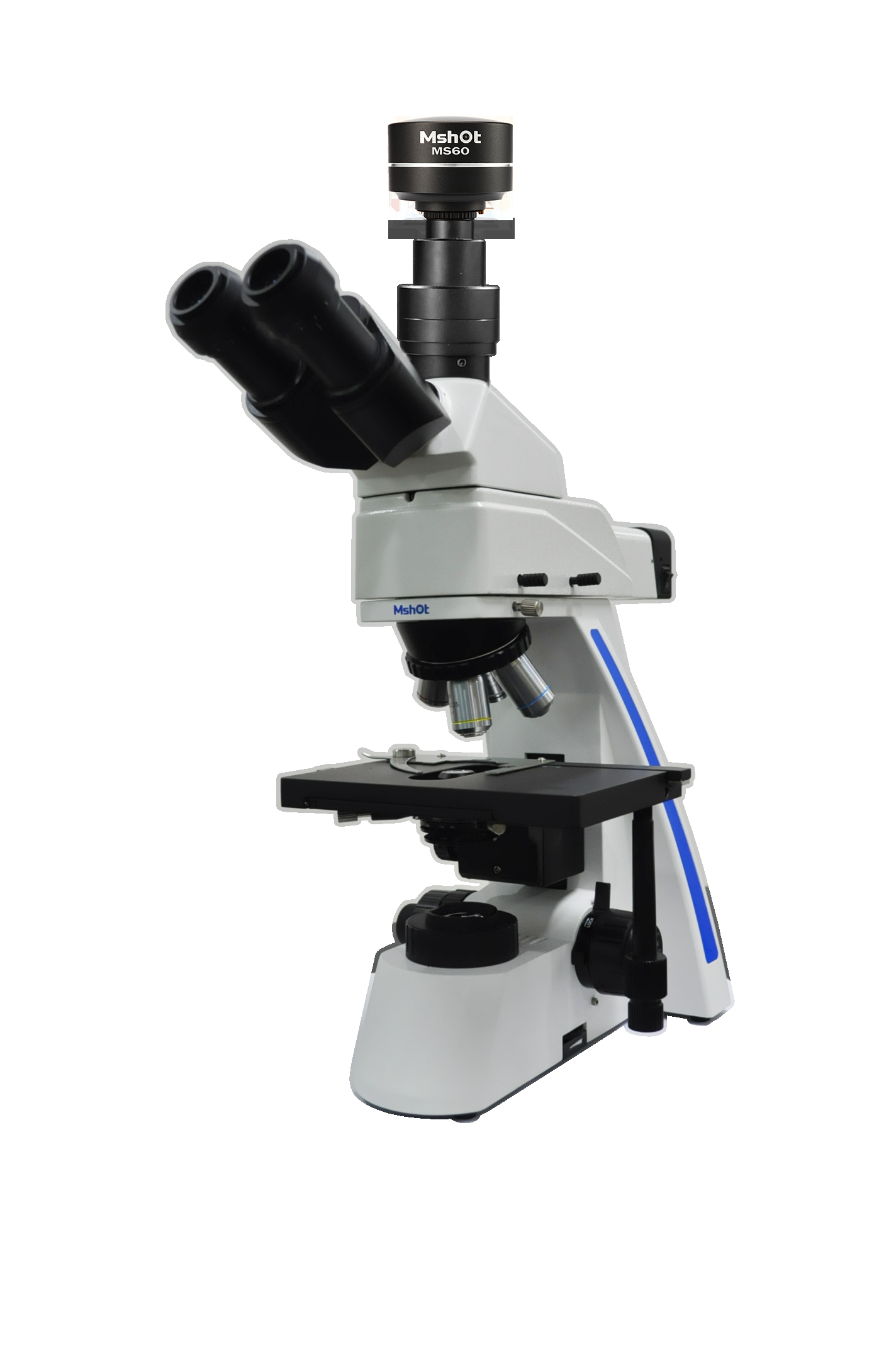
linically, pathogenic fungi can be classified into superficial fungi and deep fungi. Superficial fungi (tinea pedis) only infringe on skin, hair and finger (toe), while deep fungi can invade human skin, mucous membrane, deep tissue and viscera, and even cause systemic disseminated infection. The infection of deep fungi in the intestinal tract is characterized by fungal enteritis, which can exist independently, such as Candida infantile enteritis, or one of the manifestations of systemic fungal infection, such as AIDS with disseminated histoplasmosis. Mshot MF31-UV LED fluorescence microscope and MSX2 scientific level microscope camera is good for fungus fluorescence observation.
Direct checking under microscope is a primary diagnosis method of fungal examination, which on purpose to find mycelium and spores. Ordinary Gram staining is not specific, but fungi show character under fluorescence excitation wavelength at 380nm. Fluorescence microscope can improve specificity and bring more precise detection and judgement to doctors, and they can easily get accuracy report result for patient. At the same time, the operation is convenient and easy to learn.
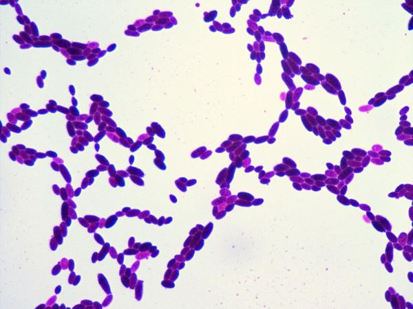
Fungus gram stained under bright field microscope 100X objective
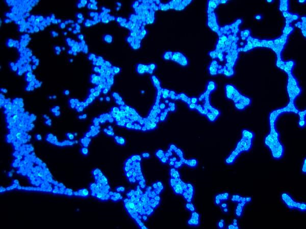
Skin Fungus under LED fluorescence microscope 40X objective excited by UV
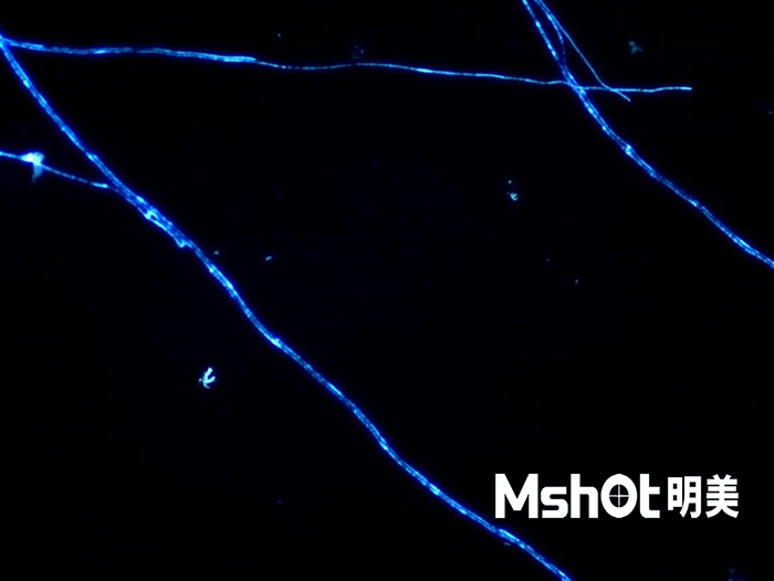
Skin Fungus Hypha under led fluorescence microscope