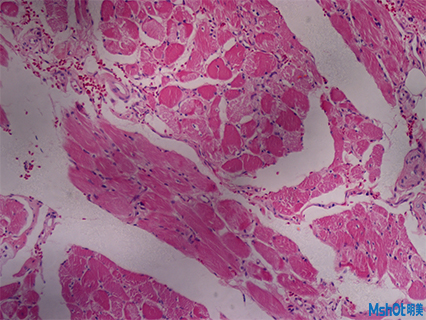
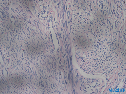
The pathological sections were made by histopathological method. The pathological section is transparent, and the morphology of tissue can be observed under the microscope. With the microscope camera, the morphological effect pictures can be preserved, compared, spliced and other operations, and the pathological diagnosis can be made through the occurrence and development of the disease.

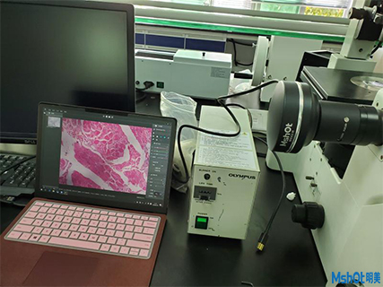
A medical technology company in Shenzhen has an Olympus inverted microscope IX71, which needs to be equipped with a set of high-pixel microscope camera to take pathological sections and fluorescent sections. According to the actual needs of teachers, Shenzhen regional engineers recommended MSHOT scientific research grade microscope camera MSX2. This microscope camera uses large target high-performance imaging chip, and designs USB3.0 data transmission interface, which has high resolution Its color performance is an ideal tool for liquid based cell analysis, immunohistochemistry, bone marrow cell analysis and other pathological diagnosis with high color requirements. In addition, it is also suitable for bright and dark field, phase contrast, polarized light, DIC, fluorescence imaging and other fields.

