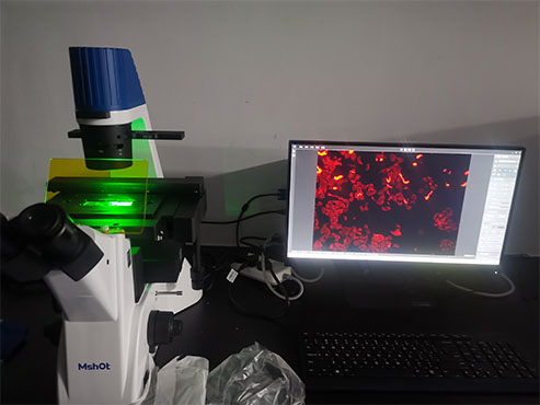
We often need to label antibodies with enzymes or fluorescent dyes. Tissue or cells are incubated with unlabeled two specific primary antibodies (from different species). After washing off the excess primary antibody, use two different fluorescent dyes to label the secondary antibody and incubate the tissue or cells respectively. After washing off the excess secondary antibody, the two antigens can be localized and quantitatively observed. The main tool is a fluorescence microscope with two corresponding excitation filters.

The equipment used in this experiment is an inverted fluorescence microscope MF52-N with a 12.5-megapixel microscope camera MSX2. The user can better record the marks of the sample. The customer is satisfied with the effect of the software processing and purchases it directly.
MSHOT inverted fluorescence microscope MF52-N is used for fluorescence microscopic observation in the fields of biopharmaceuticals, medical detection, disease prevention, etc. It is mainly used for microscopic observation of cell tissue, transparent liquid tissue and other samples.

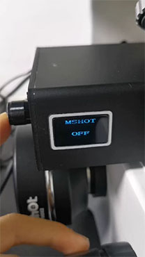
It comes with digital fluorescent illuminator, it adopts three-color three-channel design, supporting digital display design, which can realize the observation requirements of three-color fluorescence, users can choose V/B/G/Y/ The R excitation light band meets the requirements of quantitative experiments and becomes more controllable for brightness adjustment.

If you would like to get more information, please visit www.m-shot.com.