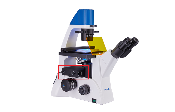
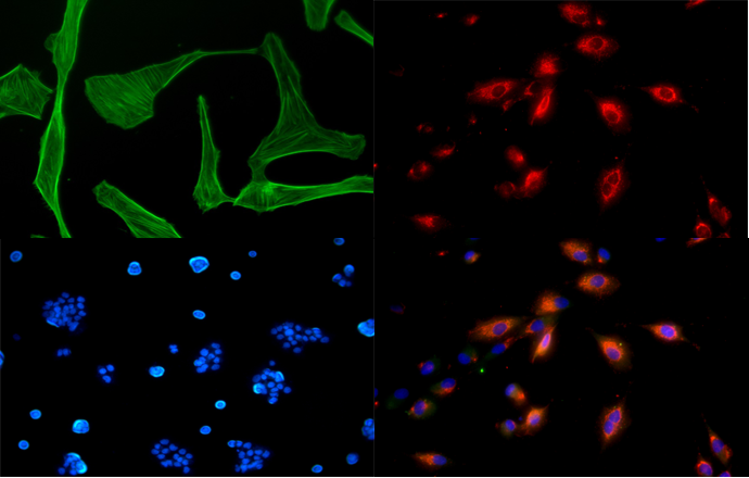
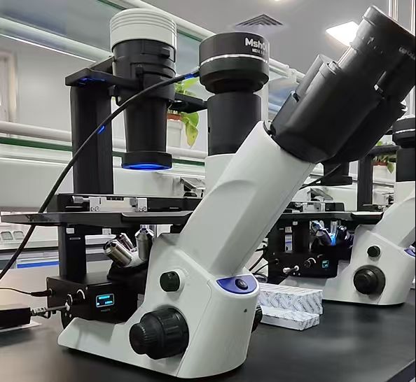
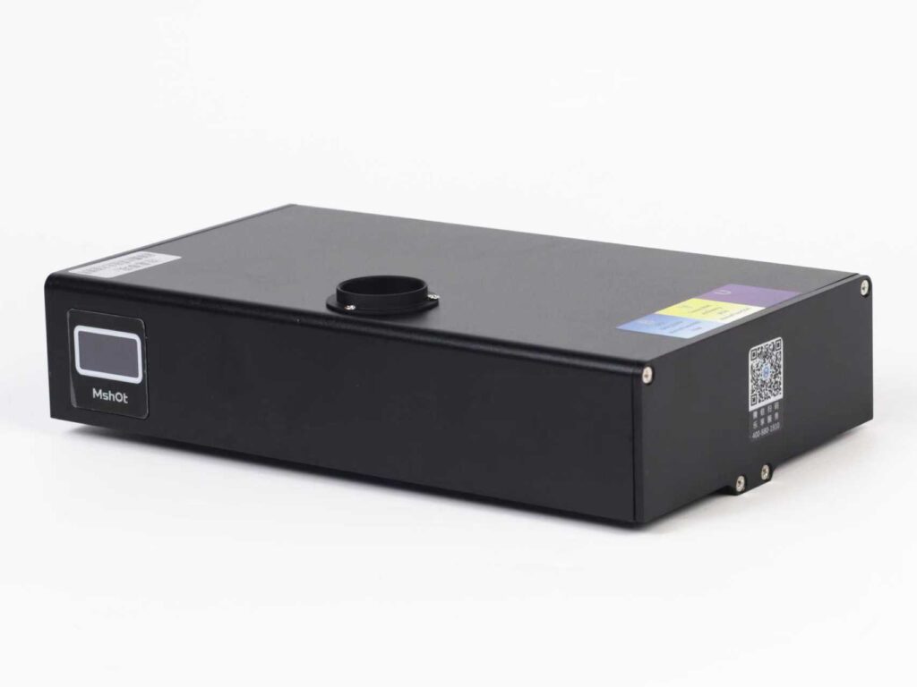
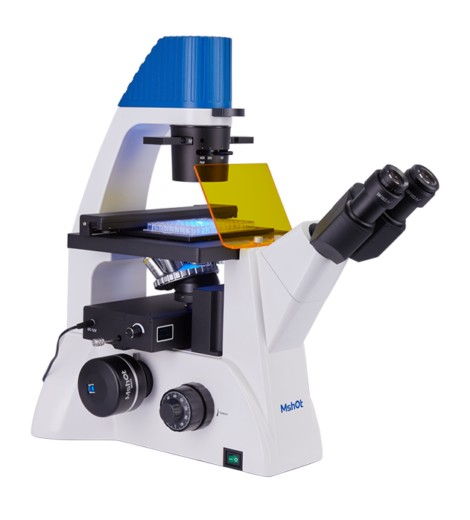
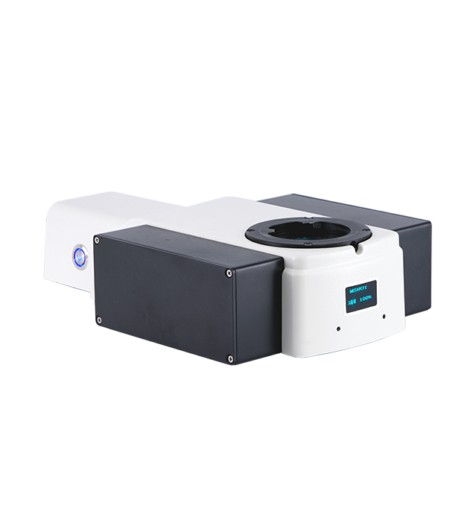
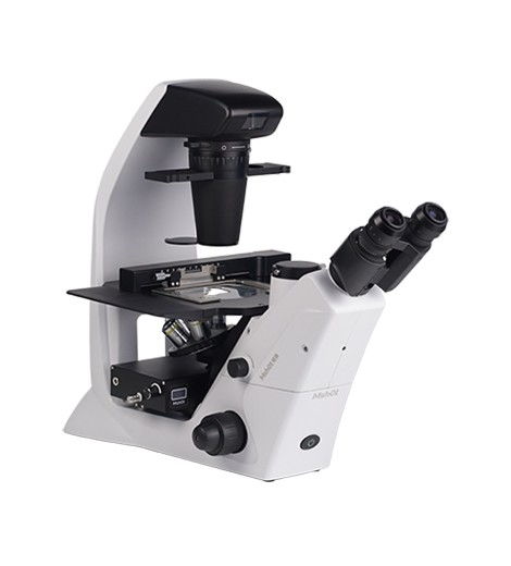
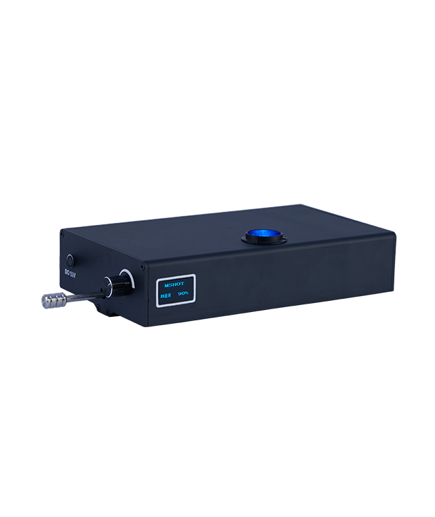
MIF-LED Inverted Digital Display LED Fluorescence Module

MIF-LED is a fluorescence attachment in LED light source, it is used to upgrade inverted biological microscope to fluorescence microscope. Special with built-in color LED lamps and fluorescence filters, the illuminator is well fit to Olympus inverted microscope for live cell. On purpose of user friendly , the products is lightweight and small volume. Digital screen shows and remember each channel light intensity from 0~100%.

Share:

Get Exclusive Offers and Updates!

    Description

    The inverted digital display fluorescence module is designed to upgrade inverted biological microscope to fluorescence function. It takes three-color four-channel structure design, with three large-viewing field fluorescence channels and one bright field channel as standard. The band switching is stable and smooth. Users can freely choose the fluorescence band and quantity according to their needs. Based on the design concept of simple appearance and easy operation, it adopts LED cold light source, integrating the driving power supply, LED excitation light source and fluorescence filter group.

    Feature

    MIF-LED inverted fluorescence module features

    ■ Compact design contains light source and filters in one unit.
    ■ Instant on-off, no need waiting of pre-heating or cooling.
    ■ Freely select fluorescence bands and quantities according to needs.
    ■ Fluorescence channel and brightness display function, display working status.
    ■ Brightness memory function, can remember the last used brightness.
    ■ Digital screen shows light intensity 0~100%
    ■ Light source synchronous switching with filter groups.
    ■ No requirement of external or added power supply.
    ■ CE, FCC, EMC, EU, ISO certified.

    MIF-LED inverted fluorescence module classic applications

    ■ Live cells culture
    ■ Chromosome analysis in cytogenetics
    ■ Histochemistry in neural tissue and localization of proteins and nucleic acids
    ■ Location and quantification of certain substances in tissues and cells
    ■ Can measure the content of some components such as protein, DNA, RNA, etc. in cells

    Detail:

    Specifications

    Item Specifications
    Filter Cube: B FB-B-M : EX: 470/40nm; DM: 505nm; EM: 530/40nm;
    FL-B-M : EX: 460-490nm; DM: 500nm; EM: 520nm LP;
    Filter Cube: G FB-G-M : EX: 530/40nm; DM: 565nm; EM: 605/55nm;
    FL-G-M : EX: 510-550nm; DM: 570nm; EM: 590nm LP;
    Filter Cube: U FB-U-M : EX: 375/28nm; DM: 415nm; EM: 460/50nm;
    FL-G-M : EX: 330-380nm; DM: 400nm; EM: 420nm LP;
    Filter Cube: Y FL-Y-M : EX: 540-580nm; DM: 600nm; EM: 610 nm LP
    Excitation Light Source 3W LED light source (5W for Y/G), adjustable brightness, visual digital display
    Observation Method Fluorescence / Brightfield switching
    Power Input  DC 12V 2A

    Resources

    You may also like…