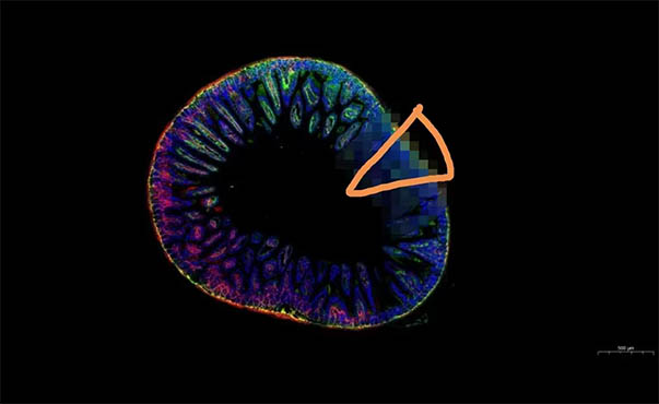
Bacterial cells are about 1 micron and invisible to the naked eye. Fluorescence microscopy enables visualization of different parts and aspects of bacteria, including the nucleus, cell membranes, organelles, and even specific proteins.

Fluorescence microscopy is often used to visualize nuclei stained with DAPI. This is a DNA stain that looks blue under the microscope. Enables visualization of pathogenic and non-pathogenic interactions between host and bacteria.
MSHOT inverted fluorescence microscope MF52-N
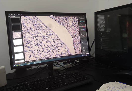
This time, MSHOT inverted fluorescence microscope MF52-N enables visualization of pathogenic and non-pathogenic interactions between host and bacteria. Inverted fluorescence microscope MF52-N is often used in the cell room to observe the fluorescent staining of cells for analysis and research. It can be used for pathological research in hospitals; it can also be used for paper publication, etc.

The inverted fluorescence microscope MF52-N is equipped with a long working distance plan objective lens and a large field eyepiece, and the image is clear. The epi-fluorescence microscope system adopts a modular design concept, which can quickly adjust the illumination system and switch the fluorescence filter assembly.

Inverted fluorescence microscope MF52-N is used for microscopic observation of cell tissue, transparent liquid tissue, or fluorescence microscopic observation in the fields of biopharmaceuticals, medical detection, disease prevention, etc.
If you would like to get more information, you could visit www.m-shot.com.