In microscopy, fluorescence refers to the phenomenon of photoluminescence. The physical phenomenon that certain molecules can absorb light of a certain wavelength and then emit light of other wavelengths. Such molecules are generally called fluorophores. Typically, due to the Stokes shift, there is a difference between the maximum excitation and emission spectra of a fluorophore, the energy of the emitted light is lower than the energy of the absorbed light, and the wavelength of the emitted light is longer than the excitation light.
There are two types of fluorescence phenomena: autofluorescence and secondary fluorescence. Autofluorescence, also known as intrinsic fluorescence, means that it can emit fluorescence directly after being irradiated; secondary fluorescence means that people need to use fluorescent dyes to mark substances that cannot or only partially emit weak fluorescence, and then can be excited by incident light. Fluorescence. At present, in fluorescence microscopy, secondary fluorescence is mainly used.
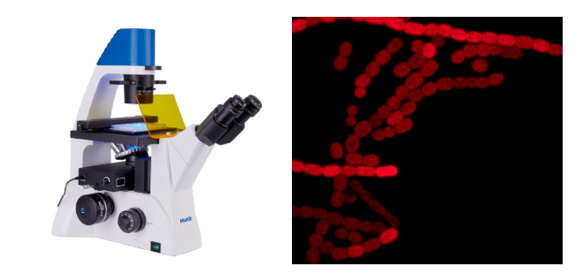
Autofluorescence of Nostoc chloroplasts taken by Mshot MF52-N
(1) Excitation light source: mainly includes the following two indicators
The light source spectrum covers the emission spectrum: fluorophores are maximally excited in a specific wavelength range, so the spectral range of the excitation light source should match the excitation characteristics of the fluorophore in the sample being observed.
The intensity of the emission spectrum band: When the fluorescence signal is weak, the user needs to use a higher-brightness excitation light to generate a stronger emission fluorescence signal.
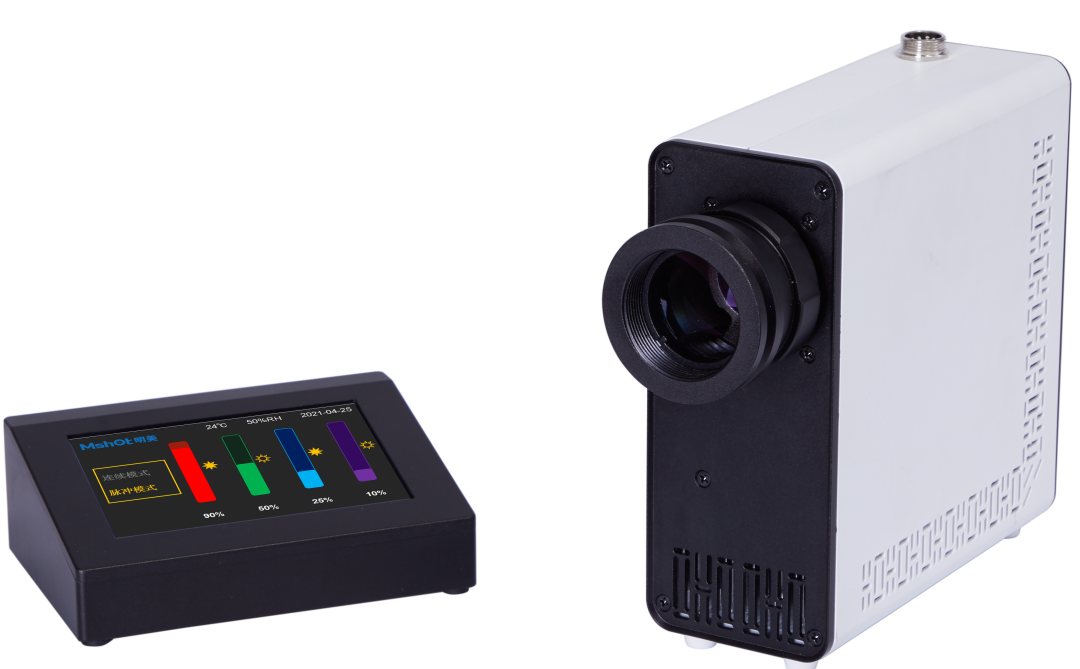
Four-channel LED light source MG120 Wide spectrum + high power
(2) Excitation block: a set of filters used to form specific excitation/emission, generally including the following three:
Excitation Filter EX: Filter specific wavelengths of light in the light source to excite specific dyes in the sample.
Emission filter EM: Allows specific wavelengths of light emitted by the fluorophore to pass.
Dichroic mirror DM: Reflects the light in the excitation band and transmits the light in the emission band. Transmission fluorescence microscopes do not have this filter, but the current mainstream is epi-fluorescence microscopes, which have dichroic mirrors.
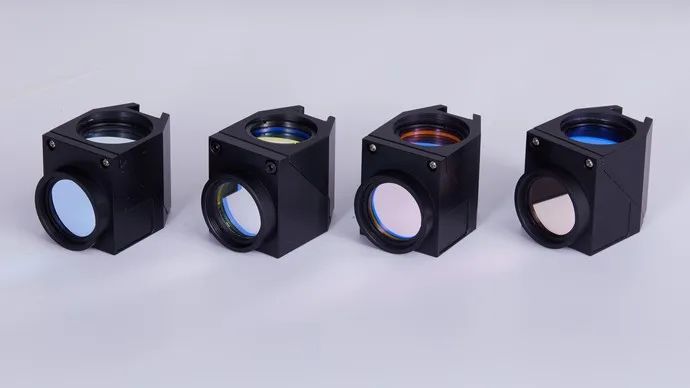
Mshot customize excitation blocks for different frequency bands for Olympus, Nikon, Leica, Zeiss
Fluorescence microscopy utilizes the phenomenon of autofluorescence or secondary fluorescence, and is widely used in microscopic fluorescence observation in the fields of biology (medicine), minerals, and materials science.
①In the field of biology, mainly by means of fluorescent dye labeling, cells and submicroscopic cellular components can be identified accurately and in detail.
② In the field of mineralogy, it is usually used to study substances with autofluorescence properties, such as petroleum, coal, graphene oxide and other minerals.
③ In materials science, it can be used in the textile industry or paper industry to analyze fiber-based materials, including textiles and paper, and in the field of semiconductors, it can also be used to observe materials with fluorescent properties such as magnetic beads.
Observation of SBS Modified Asphalt by Mshot MF31-LED Fluorescence Microscope
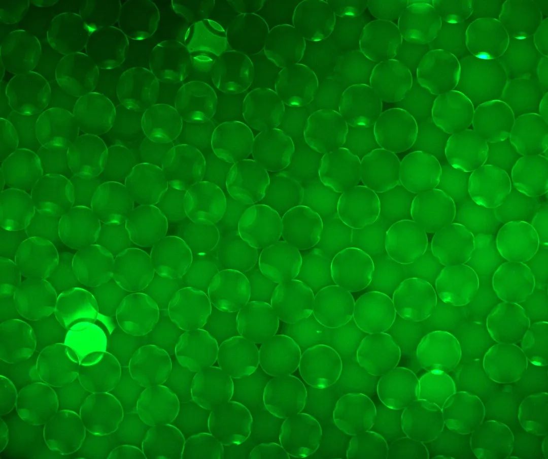
Magnetic bead fluorescence image taken by MF43-N
Fluorescence microscopy is the most widely used and most complex in the field of biology, mainly for labeling and imaging bacteria, cells, tissues or small organisms (such as zebrafish and other model organisms). Labeling at the molecular level. The selection of fluorescent dyes usually needs to consider several factors, one is the labeling position of the fluorescent dye, and the other is the corresponding excitation characteristics of the dye, including absorption spectral characteristics and emission spectral characteristics. With the help of fluorescent dyes, fluorescence microscopy is not limited to proteins, it can also stain nucleic acids, glycans, and even non-biological substances such as calcium ions can be detected. The following lists some common fluorescent dyes and their applications according to the position of dye binding:
![PM}4C(1BIP(ZGJ]I{CI7TU3.png PM}4C(1BIP(ZGJ]I{CI7TU3.png](https://m-shot.com/upload/ueditor/20220709/202207091628465831.png)
FITC fluorescent staining taken from MF52-N
(1) Immunofluorescence (IF): Mainly labeled proteins. Immunofluorescence technology utilizes the characteristic that antibodies can specifically bind to corresponding antigens. There are two main ways: one is to use endogenous fluorescent signals, that is, to genetically link fluorescent proteins to target proteins through cloning technology, such as GFP, etc.; two It uses fluorescently labeled antibodies to specifically bind to the target protein.
FITC (fluorescein isothiocyanate): is an organic fluorescent dye with excitation/emission peaks at 495/517 nm, which can be combined with groups on proteins, mainly used in immunofluorescence and flow cytometry.
TRITC (tetramethylrhodamine-5(6)-isothiocyanate): a derivative belonging to the rhodamine family, the 550/573 nm excitation/emission peak of this dye can be combined with proteins.
Huaqing dyes (cy3, cy3.5, cy5, cy5.5, cy7, etc.): less non-specific adsorption, high extinction coefficient, and high fluorescence quantum yield, so that the background of the label is weak and the signal is strong. In addition to cell visualization, they are also commonly used in biological screening, western blotting, biomedical visualization, in vivo imaging in small animals, etc.
Alexa Fluor series: belongs to the series of negatively charged and hydrophilic fluorescent dyes, which are sulfonated forms of different basic fluorescent substances. These product labels cover the corresponding excitation wavelengths, and have better stability and fluorescence brightness than dyes such as FITC.
![2ZE~4S6LMK3D]J~XIPHG]]T.png 2ZE~4S6LMK3D]J~XIPHG]]T.png](https://m-shot.com/upload/ueditor/20220709/202207091634511097.png)
DAPI fluorescent staining taken from MF52-N
(2) DNA staining: It mainly marks nucleic acids. Like proteins, labeling and researching nucleic acids is also of great significance. For example, staining and labeling the nucleus (the main component of which is nucleic acid) can determine the number and location of cells.
DAPI (4',6-diamidino-2-phenylindole): When DAPI binds to double-stranded DNA, it has a maximum absorption peak at 358nm and an emission maximum peak at 461nm. The wavelength range covers blue to turquoise. DAPI also binds to RNA, but does not produce as much fluorescence as it does to DNA. DAPI can penetrate the intact cell membrane, so it is widely used for the staining of living and fixed cells.
Hoechst (33258, 33342, 34580): All three dyes can be excited by ultraviolet light around 350nm and emit blue indigo fluorescence at 461nm. Similar to DAPI, Hoechst penetrates intact cell membranes and is widely used for live and fixed cell staining.
PI (Propidium iodide): is a DNA stain that cannot penetrate cell membranes. After binding to nucleic acid, the maximum excitation wavelength is 538nm and the maximum emission wavelength is 617nm. Because the stain cannot enter intact cells, this stain is often used to distinguish live from dead cells in a population of cells.
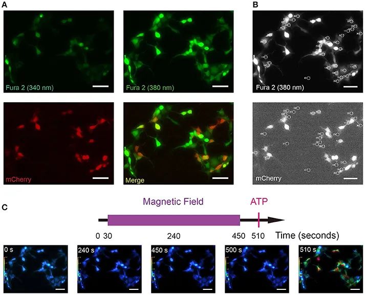
Image of research with Fura2 calcium ion probe quoted from public literature
Front. Neural Circuits 11:11. doi: 10.3389/fncir.2017.00011
(3) Ion imaging: Studying the flow and distribution of various ions in cells plays an important role in the in-depth understanding of cell activities. Various ion indicators, such as calcium ion, sodium ion, magnesium ion, zinc ion and other indicators, are combined with ions to form fluorophores, and the spectral characteristics are used to detect the distribution state of the corresponding ions.
In the process of applying fluorescence microscopy for imaging, not only the quality of imaging, including resolution, contrast, fluorescence signal strength, background light, multi-channel imaging, etc., but also the influence of fluorescent dyes and illumination on the sample must be considered. Such as fluorescent dye toxicity, phototoxicity, photobleaching, etc. The following lists common challenges and solutions in the application of fluorescence microscopy:
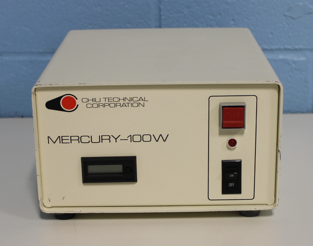
Mercury lamps require complex control boxes with timers
(1) Selection and use of excitation light source. Traditional fluorescence imaging uses a high-pressure mercury lamp, which has specific spectral characteristics and high light intensity, but there are many troubles to use. First, it needs to be warmed up, and then the light intensity is adjusted by ND filters, and the final lifetime may be as short as 200 Hours, there will be a problem of light intensity attenuation before the end of life, you need to adjust the ND filter, you need to re-calibrate after replacing the bulb. At present, many applications use LED fluorescent light sources to replace mercury lamps as fluorescent light sources, such as wide-spectrum high-power LED fluorescent light source MG-100, which has a long life and can hold up to 50 mercury lamps. The light intensity is more stable and controllable, and it can be used immediately. , save trouble and worry.
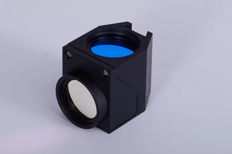
Mshot customizable filter set for Olympus, Nikon, Leica, Zeiss
(2) Filter quality and matching. The quality of the filters in the excitation block can affect the transmittance of excitation and emission light, affect fluorescence efficiency, and cause stray light and background noise problems. At the same time, whether the parameters of the filter can match the excitation and emission peaks of the dye will also have a significant impact on the fluorescence effect. For simple fluorescence needs, Mshot recommends using a digital display fluorescence module, which integrates common excitation blocks such as light source and BGU, and can upgrade fluorescence observation for the four major brands of microscopes. It is easy to use and has good effects. For professional fluorescence needs, Mshot provides excitation boxes and filter sets that match the four major brands of mainstream fluorescence microscopes. Different filter combinations can be customized on demand. For example, the calcium ion Fura2 probe needs to be excited by U and G emission. The B emission filter set is not suitable.
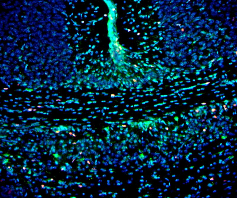
Fluorescence images of mouse brain slices taken by MF43-N+MC50-S
(3) Phototoxicity and fluorescent dye toxicity: In fluorescence imaging in the field of biology, the effects of phototoxicity and fluorescent dye toxicity on samples should be considered, such as organic molecules in cells that absorb light and decompose when they react with oxygen to generate free radicals. Some fluorescent dyes themselves can also generate free radicals, which are the main sources of damage to biological samples such as cells. The usual coping ideas are: ① Use fluorescent dyes with lower energy (longer wavelength) excitation spectrum; ② As low light intensity and short excitation time as possible, it is necessary to maintain a balance between imaging quality and sample health; ③ Reduce the frame rate, especially in long-term fluorescence imaging, in this case, it is necessary to improve the sensitivity of the camera to ensure the capture of weak fluorescence signals.

Single-channel fluorescence image-- Multi-color fluorescence image synthesized by software
(4) Multicolor fluorescence imaging: it is widely used in the field of biology, especially when imaging live cells, it is necessary to label multiple substances at the same time and display them in one image, while most fluorescent dyes have broad emission spectrum, fluorescence spectrum overlap occurs when multiple dyes are used for labeling. For multi-color fluorescence imaging, there are two ways to deal with it. One is to use the corresponding single-channel narrowband filter for each dye, and finally to synthesize it through software. This method puts forward higher requirements for imaging speed and software image processing; It uses a wide-spectrum light source for simultaneous excitation, and selects a double-pass filter or a multi-pass filter. The characteristics of this filter are that it allows two or more wavelengths of fluorescent signals to pass through. One or more fluorescent dyes are required in this way, and the adjacent emission spectral ranges do not overlap, and the obtained fluorescent signals can be better distinguished.
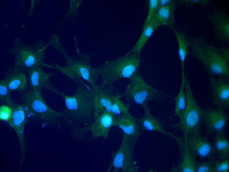
Fluorescence shooting with double-channnel filter, B and G fluorescence signals can be observed simultaneously under the microscope
![O]K(A8WTHK$Z7]]LF_}]MT1.png O]K(A8WTHK$Z7]]LF_}]MT1.png](https://m-shot.com/upload/ueditor/20220709/202207091648402417.png)
Recommended: MF23/MF31+ ordinary microscope camera MSX1/MC50-S/MS60/MD50 and other digital display LED fluorescence modules, customizable monochromatic or trichromatic excitation, push-pull switching, out-of-the-box high numerical aperture fluorescence objective lens, high definition Digital pictures can be taken with high fluorescence transmittance and high fluorescence transmittance, which is convenient for issuing reports, and multi-color fluorescence images can be synthesized.
![S6Y9JT8J_1VVJ%H4S]_UO27.png S6Y9JT8J_1VVJ%H4S]_UO27.png](https://m-shot.com/upload/ueditor/20220709/202207091649119198.png)
Recommended: MF53-N/MF43-N + MG100/MG120 + high sensitivity camera MC50-S/MS23/MSH12 .
research grade fluorescence microscope body, with better fluorescence effect and stronger expansion performance, to meet various needs.
Sixtuble turntable fluorescence attachment, you can choose the excitation block according to your needs, and realize the observation of various fluorescent dyes.
Special filter sets such as double-pass can be customized to achieve more efficient observation applications such as FISH.
LED excitation light source, high-power wide-spectrum excitation effect, easy to use, long life and cost-effective.
High-sensitivity camera, more efficient real-time feedback of dynamic images, with software to achieve FISH and other applications.
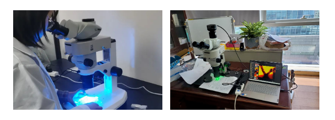
Recommended: MZX81/MZX100 + ordinary microscope camera MSX1/MC50S/MS60/MD50 etc.
High-quality plan (apochromatic) objective lens, independent parallel dual optical paths, large zoom ratio, good imaging effect.
Digital display fluorescence module, optional dual-color excitation such as BG, and UV separately external to reduce damage to samples and internal coating.
Widely used in zebrafish, nematodes, Drosophila, Sequence inspection of the formation of text and printed text, new materials and other fields, rich experience.
![]ETG0IR%BMRHA6WWWD2Z(3C.png ]ETG0IR%BMRHA6WWWD2Z(3C.png](https://m-shot.com/upload/ueditor/20220709/202207091650151435.png)
Recommended: digital display fluorescent illuminator, or batch custom fluorescent illuminator
Compatible with most of the four major brands of infinity optical microscopes, cost-effective upgrade fluorescence observation
The digital display screen can directly display the current band and brightness, which is convenient for quantitative analysis.
Built-in LED fluorescent light source, optional monochromatic or three-color excitation such as BGU, optional motorized switching or four-color excitation.
![RB%FUP)FF4~GY`YJ91]NQC6.png RB%FUP)FF4~GY`YJ91]NQC6.png](https://m-shot.com/upload/ueditor/20220709/202207091650385479.png)
Recommended: Broad-spectrum high-power fluorescent light source MG-120, four-channel light source MG-120
It can match the four major brands of mainstream fluorescence microscopes, covering the visible light excitation spectrum, and the excitation effect is stable.
Touch screen controller, intuitive and easy-to-use operation experience, can add intelligent functions such as light off when people walk away.
Long life, ready to use, 1 LED light source tops 50 mercury lamps, no need to preheat.
Light intensity adjustment is highly linear, MG-120 supports software trigger and TTL level pulse mode trigger.
Micro-shot is a high-tech enterprise focusing on the research and development and sales of microscopic imaging products. We have always insisted on operating with integrity, serving with heart, and providing microscopic imaging solutions. Provide efficient and stable products and services for our customers in education, scientific research, medicine, and industrial testing. We always focuses on customer needs, takes quality as the foundation, and takes innovation as the direction to promote the digital, electric and intelligent process of the microscopy industry.
If you would like to get more information, you could visit https://www.m-shot.com.