Olympus CKX53 upgrades to multi-channel fluorescence

Olympus CKX53 is a compact inverted biological microscope for cell culture, as the start product of Olympus, it gives stable performance and a comfortable workflow for a variety of cell culture needs, including live cell observation, cell sampling and handling, image capture, and fluorescence observation. The upgradable base configuration contains a bright field, phase contrast, and fluorescence. 

The original Olympus CKX53 fluorescence accessories include a 100W mercury lamp and a fluorescent illuminator. There is a large mercury lamp light box installed behind the bracket, which requires debugging and installation by an experienced microscope engineer.

What is the replacement solution for mercury illuminator?

Why replace the mercury fluorescence illuminator?

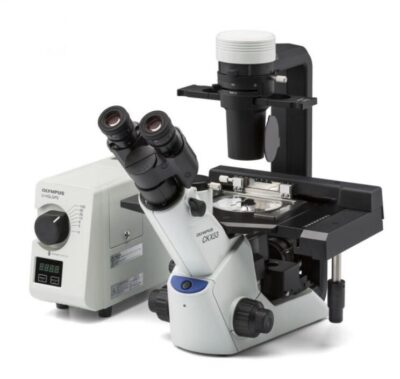
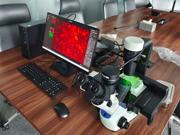
Mercury lamp light box installation and debugging are complicated, and mercury lamps need to be replaced every few hundred hours. Each use requires preheating and cooling, anti-scalding, etc., which is inconvenient for users and sellers are not tired of maintenance. In particular, mercury element pollution to the environment, human body toxicity, and the inability to separate ultraviolet light are dangerous to users. Therefore, mercury lamp lighting is gradually discontinued. Users need to find third-party fluorescent accessories.
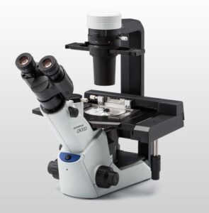
Mercury lamp houseMercury lamp house

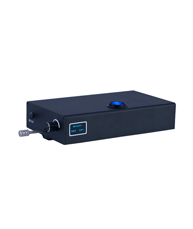
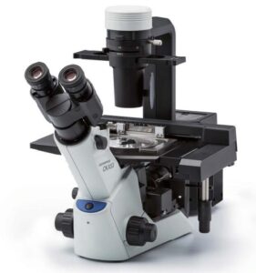
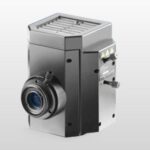
Install at the back of the microscope frame


Mercury fluorescence package of Olympus CKX53

Olympus CKX53 mercury fluorescence package

LED fluorescence module for Olympus CKX53 microscope
Olympus CKX53 with MIF-LED fluorescence module

Olympus CKX53 with LED fluorescence module

Why is an LED fluorescence module a good choice for Olympus CKX53?

Convenient installation

  • The modular design is compact and lightweight, and the module is about 2KGS
  • Easy to install, users do not need special training and can install according to the diagram
  • No need to make any changes to the Olympus CKX53 parts, or add other accessories, all included

Easy and safe to use

  • The optical path center is pre-adjusted and ready to use, and no debugging is required
    LED lamp beads are cold light sources, no need to preheat and cool down.
  • The illuminator and fluorescence excitation block and filter are integrated into one and switch to any fluorescence channel in one step
  • CE and ISO certification, Evident registered third-party supplier to ensure quality

Budget saving, users like it, sellers worry-free

  • The production cost of LED lamp beads is much better than mercury lamps, and the purchase cost is less
  • 50,000 hours of lighting life, low maintenance cost
  • 3W~6W lamp beads, 12V2A power consumption saves power
  • Simple structure, detachable internal structure, easy maintenance for sellers

Product advantages

  • The supported excitation band includes 330nm~625nm
    The supported fluorophores include common FITC, PI, TeaxsRed, mCherry, Cy3, Cy5, DAPI, etc.
  • The OLED digital display screen displays the brightness of each channel from 0 to 100% simultaneously, which is convenient for fluorescence quantification
  • The memory function displays the brightness of the last election by default, and the brightness of each channel is independently memorized
  • One module supports up to 4 fluorescence channels at a time

Introduction of the LED fluorescence module

MIF-LED is a fluorescence attachment in an LED light source. It is used to upgrade an inverted biological microscope to a fluorescence microscope. The illuminator is wel suited to an Olympus inverted microscope for live cells, especially with built-in color LED lamps and fluorescence filters. The product is lightweight and small in volume to be user-friendly. The digital screen shows and remembers each channel’s light intensity from 0% to 100%.

Olympus CKX53 LED fluorescence installation

Olympus CKX53 with LED fluorescence module installation photos

Fluorescence observation of live cells


Standard configuration
ModelLED lampFilter wavelengthMainly Applied Fluorochrome
Excitation filterDichroic mirrorEmission filter
BBlue475/30nm>505nm530/40nmGFP / FITC /EGFP/ Malaria diagnostic/ Alexa 488 / Cy2@ / Fluo-4 / FluorX@ / Fluoro-Jade
GGreen530/40nm>570nm575nmLPPI / EB / EH /TRITC
YYellow560/40nm>600nm610nmLPmCherry / Texas Red /     AlexaFluor 594
UUV375/30nm>415nm460/50nmDAPI / Hoechst 33342&33258 / AMCA/AMCA-X / Alexa 350
Remark*Olympus CX41 UV excitation only can be long-pass filter
Light sourceBlue &UV: 3W LED cold lamp for each filter group
Green/Yellow: 5W LED cold lamp for each filter group
ObservationFluorescence
Bright field & phase contrast by microscope original lighting
OperationLever : B, G, UV/O
Power controlRota-table knob, continuously adjustable brightness
ScreenDigital screen to show light intensity 0~100%, rememer light intensity of each color
Input powerDC 12V 2A
ShellHigh rigid precision-cast aluminium with coating and vents
Live cell DAPI stain
cells dyes of green excitation

Stained cell imaging performance under the LED fluorescence module upgraded inverted microscope